
Last updated: June 17, 2026
Most spine MRI reports look terrifying on first read — and most of them shouldn't be. In peer-reviewed imaging studies of pain-free adults, disc bulges, degeneration, and even small herniations show up in a striking proportion of healthy people, especially over the age of 40 (Brinjikji et al., AJNR, 2015). That single fact changes how you should approach your own report. I'm Omer Boshara, a board-certified, DWG-certified spine surgeon based in Stolberg, Germany, and I review spine MRIs for second opinions from patients all over the world. This guide on how to read a spine MRI report is written to help you understand the language, separate signal from noise, and ask better questions before you ever consider surgery.
Quick Answer
To read a spine MRI report, focus on three things: the level (e.g., L4-L5), the finding (bulge, protrusion, extrusion, stenosis, modic changes), and whether the finding contacts or compresses a nerve root or the spinal cord. The most important rule: imaging findings only matter when they match your symptoms. Many "abnormal" findings are normal age-related changes and do not require surgery.
Key Takeaways
- A spine MRI report has predictable sections: clinical history, technique, findings (level by level), and impression.
- "Disc bulge," "desiccation," and mild "facet arthropathy" are extremely common and often age-related, not injuries.
- A herniation is described by shape (protrusion vs extrusion) and what it touches (nerve root, thecal sac, cord).
- Stenosis means narrowing — it only matters clinically if symptoms match the level and side.
- Red flags include cord compression, cauda equina signs, infection (discitis), tumour, or fracture with neural compromise.
- Imaging-symptom mismatch is the single biggest reason patients are advised into surgery they may not need.
- An independent second opinion on the MRI itself (not just the surgical plan) is reasonable before any elective spine operation.
What Do the Different Sections of a Spine MRI Report Mean
A spine MRI report is structured into four predictable parts: clinical indication, technique, findings, and impression. Reading them in order tells you what the radiologist was looking for, how they looked, what they saw, and what they concluded.
- Clinical indication / history: Why the scan was ordered (e.g., "left leg pain, 6 weeks"). If this is wrong or incomplete, the interpretation can be skewed.
- Technique: Which sequences were used (T1, T2, STIR, sometimes with contrast). T2 images show fluid bright and are best for discs and nerves; T1 shows anatomy and fat; STIR highlights inflammation or oedema.
- Findings: A level-by-level description (e.g., L1-L2, L2-L3, etc.) of discs, vertebrae, canal, foramina, and soft tissues.
- Impression: The radiologist's summary of what matters most. This is usually where the clinically relevant findings are flagged.
Quick rule: if the impression doesn't mention a finding, it's usually considered minor.
How Do Radiologists Describe Disc Herniation on an MRI
Radiologists follow a standardised lexicon (Fardon et al., combined task force of the North American Spine Society, ASSR, and ASNR, 2014) to describe disc problems. The terminology sounds dramatic but each word has a specific, narrow meaning.
A herniation only matters when the report links it to a nerve structure: "contacts," "abuts," "displaces," or "compresses" the exiting or traversing nerve root.
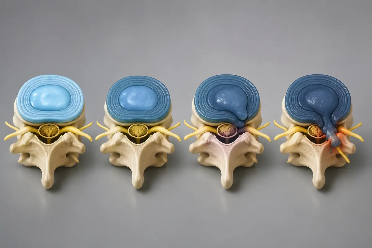
What's the Difference Between a Bulging Disc and a Herniated Disc
A bulging disc is a broad, circumferential outpouching of the disc — usually a normal part of ageing. A herniated disc is a focal displacement of disc material through a defect, and it's more likely (though not guaranteed) to cause symptoms.
- Bulge: think of a tyre that's slightly flattened all the way around. Usually does not compress a single nerve.
- Herniation (protrusion or extrusion): think of a focal blister. Can push on a specific nerve root and cause leg pain, numbness, or weakness in that nerve's distribution.
Choose to worry less if: the report says "broad-based bulge, no nerve root contact, no canal stenosis." Worry more — and seek review — if it says "extrusion with compression of the traversing L5 nerve root" and you have matching left-leg symptoms.
What Does Degenerative Disc Disease Look Like on an MRI Report
Degenerative disc disease (DDD) on an MRI report usually appears as a combination of disc desiccation (dark T2 signal), loss of disc height, annular fissures, endplate changes (Modic types), and sometimes osteophytes. Despite the name, it is not really a "disease" — it's the imaging signature of normal wear over time.
Modic changes are worth knowing:
- Modic Type 1: Oedema in the vertebral endplate. May correlate with active low back pain in some patients.
- Modic Type 2: Fatty replacement. Usually more stable.
- Modic Type 3: Sclerosis. Late-stage change.
DDD alone, without nerve compression or instability, is rarely a surgical indication.
Can a Spine MRI Show Nerve Compression
Yes — MRI is the best non-invasive test for showing nerve compression. The report will use specific language to grade it: "contact," "abutment," "displacement," "mild/moderate/severe compression," or "effacement of the thecal sac."
Look for these phrases in your report:
- "Foraminal stenosis" — narrowing where the nerve exits the spine
- "Lateral recess stenosis" — narrowing where the nerve travels before exiting
- "Central canal stenosis" — narrowing around the spinal cord or cauda equina
- "Nerve root impingement / compression" — direct pressure on a root
Critical point: the MRI shows anatomy, not pain. A nerve can look compressed and feel fine, and vice versa. This is why the next section matters more than any single phrase in your report.
What Medical Conditions Can a Spine MRI Diagnose
A spine MRI can diagnose or rule out: disc herniation, spinal stenosis, spondylolisthesis (vertebral slippage), facet joint arthropathy, vertebral fractures (acute and chronic), infections (discitis, osteomyelitis, epidural abscess), tumours (primary or metastatic), multiple sclerosis plaques in the cord, syringomyelia, and inflammatory conditions like ankylosing spondylitis.
It is less useful for: muscular pain, very early inflammatory disease (sometimes), and functional or instability problems that only show on dynamic imaging (flexion-extension X-rays may be needed).
What Are Common Red Flags in a Spine MRI Report That Suggest Serious Issues
Certain findings warrant urgent in-person review, not a leisurely second opinion. If your report mentions any of the following, contact a spine specialist promptly:
- Cord compression with T2 signal change in the spinal cord (myelomalacia)
- Cauda equina compression (especially with saddle numbness or bladder symptoms — this is an emergency)
- Discitis / osteomyelitis — signs of spinal infection
- Epidural abscess — collection compressing neural structures
- Tumour / mass lesion / suspicious metastasis
- Acute fracture with retropulsion into the canal
- Pathological fracture in a known cancer patient
These are the situations where I tell patients clearly: stop researching, get seen in person.
Who Should Get a Spine MRI and Who Might Not Need One
Major guidelines (including NICE NG59 for low back pain and sciatica) recommend against routine MRI for non-specific low back pain in the first 4-6 weeks unless red flags are present. MRI is appropriate when symptoms are persistent, neurological (weakness, numbness, radiating pain), or when surgery or injections are being considered.
You probably need an MRI if:
- Leg or arm pain in a nerve distribution lasting more than 4-6 weeks
- Progressive weakness or numbness
- Suspected infection, tumour, fracture, or inflammatory disease
- Failed conservative care and you are weighing surgical options
You probably don't need an MRI (yet) if:
- Acute low back pain under 4-6 weeks with no red flags
- Pain that is improving with movement and basic care
- No neurological symptoms
What Imaging Findings Suggest I Might Need Spine Surgery
Surgery is considered when imaging shows a structural problem that matches your symptoms and conservative care has failed — or when a red flag (cauda equina, progressive weakness, cord compression, infection, instability) requires it. Imaging alone never determines surgery.
Common scenarios where surgery is reasonably discussed:
- Persistent radicular leg pain with an MRI showing a matching nerve root compression after 6-12 weeks of proper conservative care.
- Neurogenic claudication from significant central canal stenosis affecting walking distance.
- Progressive motor weakness with a corresponding compressive lesion.
- Cauda equina syndrome (urgent).
- Instability (e.g., high-grade spondylolisthesis with symptoms).
- Tumour, infection, or unstable fracture.
If the MRI finding doesn't explain your symptoms, operating on it rarely fixes the pain. This is the single most important sentence in this entire article.
How Much Does a Spine MRI Interpretation Typically Cost
Costs vary widely by country and health system. The MRI scan itself is usually billed separately from the radiologist's interpretation, and a second-opinion review by a spine surgeon is a separate service again.
- Scan + radiology report: ranges from publicly funded (no direct cost in many European systems) to several hundred euros, pounds, or dollars privately.
- Independent surgeon second opinion (online): typically a fixed consultation fee, often a fraction of the cost — and risk — of an unnecessary operation.
I won't quote specific prices here because they shift by region and provider. Ask for a written fee in advance.
What Mistakes Do Patients Often Make When Trying to Read Their Own MRI Report
The biggest mistake is treating the report as a verdict instead of a description. The second biggest is reading the impression in isolation without matching findings to symptoms.
Common pitfalls I see weekly:
- Googling "extrusion" and assuming surgery is inevitable
- Ignoring the word "mild" and focusing on the scary noun
- Assuming a finding on the right side explains left-sided pain
- Treating a bulge at one level as the cause of pain that follows a different nerve
- Forgetting that ageing changes are normal — the same way grey hair is
How Do I Explain My Spine MRI Findings to My Doctor
Bring three things to the appointment: the report, the images themselves (on a CD or via a portal), and a written symptom map showing exactly where pain, numbness, or weakness occur. Then ask focused questions.
Useful questions to ask:
- Which finding on this MRI, if any, matches my symptoms?
- Are there findings here that are common in people without pain?
- What are my non-surgical options, and how long should I try them?
- If I had surgery, which exact finding would it target, and what's the expected benefit?
- Would you recommend an independent second opinion before deciding?
A good clinician welcomes question five.
Are There Alternative Ways to Diagnose Spine Problems Besides MRI
Yes. MRI is the most detailed for soft tissue and nerves, but it's not the only tool. The right test depends on the suspected problem.
- X-ray: bone alignment, fractures, instability (especially flexion-extension views)
- CT scan: bone detail, post-surgical hardware, fractures when MRI is contraindicated
- EMG / nerve conduction studies: functional testing of nerve injury
- Diagnostic nerve root blocks: can confirm which level is actually generating pain
- Clinical examination: still the most important diagnostic tool, and the one most often skipped
Why an Independent MRI Second Opinion Matters Before Surgery
A second opinion on the MRI itself — not just the surgical plan — catches two problems: misread imaging and imaging-symptom mismatch. Both are common, and both can lead to surgery that doesn't help. An online review by an independent spine surgeon is a reasonable, low-risk step before any elective operation.
When I review an MRI as a second opinion, I'm asking:
- Does the imaging finding actually explain the patient's pain pattern?
- Have appropriate non-surgical options been tried for an appropriate time?
- Is there a less invasive option that fits this anatomy?
- Are there red flags that change urgency?
- Would I operate on this patient in my own clinic — and if so, how?
This isn't about overriding your local surgeon. It's about making sure you walk into the decision with clear eyes.
FAQ
Is a disc bulge the same as a slipped disc?
No. "Slipped disc" is a lay term that usually refers to a herniation (protrusion or extrusion). A bulge is broader, milder, and often a normal ageing change.
Can a herniated disc heal without surgery?
Often, yes. Many disc herniations — including some large extrusions — reduce in size over months without surgery, particularly when leg pain is the main symptom and there's no major weakness.
What does "no acute findings" mean on my report?
It means the radiologist didn't see anything urgent or new — no fracture, no tumour, no infection, no severe compression. Chronic or age-related changes may still be present.
Should I worry about Modic changes?
Modic Type 1 (oedema) may correlate with active back pain in some patients. Type 2 (fatty) and Type 3 (sclerotic) are usually more stable. None automatically mean surgery.
Can MRI tell if my pain is "real"?
MRI shows structure, not pain. Real pain often exists without major MRI findings, and major MRI findings often exist without pain. The two must be correlated clinically.
How quickly should I act on a "severe stenosis" finding?
It depends on symptoms. Severe stenosis with progressive weakness, balance loss, or bladder symptoms is urgent. Severe stenosis with intermittent leg fatigue while walking is usually not an emergency, but warrants a specialist review.
Conclusion
Learning how to read a spine MRI report won't turn you into a radiologist, but it will protect you from two common mistakes: panicking over normal age-related findings, and accepting surgery for a finding that doesn't match your symptoms. Focus on level, finding, and contact with neural structures. Insist that any proposed treatment — especially surgery — be tied directly to a finding that explains your actual pain.
If you're weighing a surgical recommendation, the most useful next step is usually not more reading. It's a structured second opinion that looks at the images, the report, and your symptoms together. I offer that service online to patients worldwide, and even if you don't choose me, choose someone independent. Your spine deserves a careful decision, not a fast one.
This article is educational and does not replace personalised medical advice. For any concerning symptom — especially weakness, numbness, or bladder or bowel changes — please seek in-person evaluation promptly.